Small office. Great experience.

Address:

601 S Garfield Ave, Suite A, Traverse City, MI 49686

How Often Should I Get An Eye Exam?

The answer depends on your age. This is because age is the single biggest risk factor for eye disease, including cataracts, glaucoma, and macular degeneration. The American Academy of Ophthalmology (AAO) recommends that asymptomatic people over the age of 65 with no risk factors should have a dilated eye exam every 1-2 years. An exam like this can be performed by either an optometrist or ophthalmologist. Here in northern Michigan, we are fortunate to have a number of qualified Traverse City ophthalmologists and optometrists. For younger patients, the frequency decreases; for instance, patients 55 to 64 years old should have a dilated eye exam at least every 1-3 years.

Won’t I Know If Something Is Wrong With My Eyes?

Sometimes I am asked, “But wouldn’t I know if something was wrong with my eyes? Why do I need to have a ‘routine’ eye exam?” One word: glaucoma. Glaucoma is a potentially blinding eye disease that is insidious because it is asymptomatic until very late in the disease cycle, when much sight has been lost. This is because glaucoma causes changes in the peripheral vision that often go unnoticed because the brain can “fill in the gaps”. I recently saw a heart-breaking case of a relatively young man who thought the vision in his left eye was getting dim because of the beginning of a cataract forming; he actually has very severe glaucoma that has taken over 80% of the vision in that eye. He even told me that an eye care professional in the past had been concerned about glaucoma affecting his peripheral vision, but the patient had reassured himself by seeing if he could see his fingers in his far peripheral vision (he could). However, on formal visual field testing in my Traverse City office he demonstrated significant visual field loss, resulting in “tunnel vision”. It turns out that testing yourself with your fingers in your peripheral vision is too crude to detect glaucomatous damage.

Preglaucoma? Don’t Hesitate To Get A Second Opinion

One final word on glaucoma, because it is such a serious condition that can cause blindness. Glaucoma is a difficult disease to accurately diagnose early in the disease cycle, and a number of different tests can be necessary. With that said, some unsavory eye doctors have been known to use this fact to “overtest” patients who “might have glaucoma” or “preglaucoma”. In the majority of cases, extensive repeat testing is not necessary in glaucoma suspects or preglaucoma. My statement to all patients is that if they ever question whether or not they are being “overtested” they should simply get a second opinion, which can provide peace of mind.

Ophthalmologists Urge Eye Protection for Recreational and Professional Sports

New research shows that about 30,000 people in the U.S. go to emergency departments each year with sports-related eye injuries, a substantially higher estimate than previously reported. This April during Sports Eye Safety Month, Potthoff Eye Care & Surgery in Traverse City, MI and the American Academy of Ophthalmology remind the public that the right protective eyewear is the best defense against eye injury.

Three sports accounted for almost half of all trips to the emergency room: basketball, baseball, and air/paintball guns. Sports-related injuries can range from corneal abrasions and bruises on the lids to more serious, vision-threatening internal injuries, such as a retinal detachment and internal bleeding.

Ophthalmologists — physicians who specialize in medical and surgical eye care — continue to remind the public that most sports-related eye injuries are avoidable.

Here are some tips for both the professional athlete and the Little League star to stay safe: 

  • Athletes should wear sports eye protection that meets requirements set by appropriate organizations.
  • Parents should make sure that children wear eye protection. Most often, those who sustain sports-related eye injuries are 18 years old or younger.
  • Eye protection can weaken with age and may no longer provide adequate protection. Consider replacing when damaged or yellowed.
  • For basketball, racquet sports, soccer and field hockey, wear protective eyewear with polycarbonate lenses.
  • Athletes who wear contacts or glasses should also wear appropriate protective eyewear. Contacts offer no protection and glasses do not provide enough defense.
  • Professional athletes should also wear sports goggles that meet national standards.

“I’ve treated many patients with eye trauma because of an unintentional blow to the face,” said Rahul N. Khurana, M.D., clinical spokesperson for the American Academy of Ophthalmology. “Athletes often engage in these seemingly safe, yet rugged, high-impact sports with zero awareness about the potential risk factors. This is why eye protection is critical and can greatly reduce the number of emergency room visits treated each year.”

To learn more ways to keep your eyes healthy, visit the American Academy of Ophthalmology’s EyeSmart® website.

.

Eye doctor in Traverse City, MI tried Lumify Eye Drops

Do Lumify Eye Drops Work?

I recently wrote a post about Lumify Eye Drops, the new eyedrops made by Bausch & Lomb and marketed as having the ability to whiten and brighten a person’s eyes. I talked about the mechanism of action, type of medication involved, and safety. However, I hadn’t yet tried Lumify Eye Drops myself so I couldn’t speak to their effectiveness.

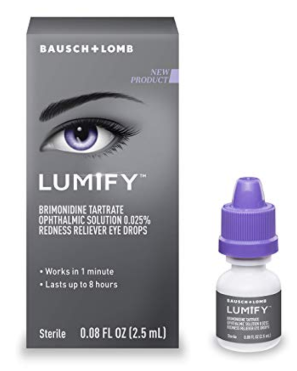
Lumify Eye drops box and bottle
Lumify Eye Drops

Well, I decided to try them when I realized that my wife had bought a bottle! Let me first say that I am quite the skeptic when it comes to new medications rolled out and marketed by big drug companies, especially when it comes to some type of cosmetic use. With that said, I couldn’t believe how well they worked! I tried it in one eye only so that my other eye could serve as a control, and within a minute or two sure enough the eye that got the Lumify drop was considerably more white! I was pleasantly surprised that it actually lived up to its claims. I ended up putting a drop in the other eye and watching it whiten in the mirror over a minute or two. It seriously works. My eyes did feel just a tad irritated over the next little bit, but not too bad. The effect lasted an hour or two by my unscientific estimate.

So yes, I can testify that Lumify Eye Drops do work. As an aside, Basuch & Lomb is probably going to make a ton of money off of Lumify. With that said, I haven’t used the drops again because I’m not particularly bothered by what little redness I have in my eyes at baseline. However, I could see using the Lumify every now and then before going out or knowing my picture is going to be taken at an event, etc. For people more bothered by chronically red eyes I could see the appeal in using Lumify more often.

Stephen Curry’s Eye Condition: What Is Keratoconus?

What is keratoconus?

Steph Curry, the star of the NBA’s Golden State Warriors basketball team recently told the press that he has keratoconus. Keratoconus is a progressive eye disease that involves the cornea, the very front part of the eye that light rays first pass through when entering the eye (it’s the part that really hurts if you scratch it and get a corneal abrasion). Corneas are ideally round or spherical, but in keratoconus there is progressive thinning in certain areas of the cornea that cause the cornea to weaken and lose its spherical shape. This results in light not being focused as well and causes blurry vision. The only way to know if you have keratoconus is to visit an eye doctor.

Much has been made in the media since Steph Curry’s announcement that he has keratoconus, but I think some perspective is in order. Like most diseases there is an entire spectrum of pathology. Severe keratoconus is extremely visually debilitating, and often requires a corneal transplant to fix. At the other end of the spectrum is mild or early keratoconus, which I suspect better describes Steph’s eye condition. I say this because he simply wouldn’t be able to see, let alone play NBA basketball if he had untreated keratoconus that was anything but mild. I certainly don’t mean to make light of his eye condition as it is indeed serious and progressive, but the claims that he became one of the best three-point shooters in history while not being able to see are a bit overblown.

How is keratoconus treated?

Steph Curry serves as a good example that in early keratoconus the vision can often be improved with contact lenses. The contacts may either be your standard soft contact lenses or hard contacts, also known as rigid gas permeables (RGPs). Contacts ride on the cornea and serve to mask it’s irregular shape, thus creating sharp vision. Historically, when a keratoconus patient could no longer achieve acceptable visual acuity with contact lenses, a corneal transplant was required. Fortunately, today we have corneal collagen cross-linking (CXL) to help stabilize and possibly reverse keratoconus before a patient requires a corneal transplant. CXL works by saturating the cornea with a chemical (riboflavin) that is then activated with ultraviolet light; the end result is the cornea becomes “stiffer” and less prone to progressively lose its shape. CXL has been used for years in Europe with great results, and was recently approved by the FDA for use here in the US. CXL will no doubt save many patients from requiring a corneal transplant.

Can I have LASIK if I have keratoconus?

No. Keratoconus is a contraindication to having LASIK because the cornea is already weak, and during LASIK corneal tissue is lasered away to bring the eye into focus without glasses. By removing corneal tissue LASIK weakens the cornea. This isn’t an issue for the vast majority of people, but in patients with keratoconus it is a bad idea to further weaken any already weak cornea. This could lead to post-LASIK ectasia, which is similar to keratoconus as the cornea starts to lose its spherical shape and causes blurred vision. With all that said, it may still be possible to have refractive surgery (so you don’t need glasses) if you have keratoconus. Some eye surgeons are performing corneal collagen cross-linking prior to PRK (similar to LASIK but with less weakening) in the right candidates.

Lumify Eye Drops

Last year Bausch & Lomb released Lumify eye drops, marketed to help reduce eye redness and give the appearance of whiter and “brighter” eyes. Lumify is available over-the-counter so they do not require a prescription from your eye doctor. Here in Traverse City, MI I have seen them on the shelves at Rite Aid and Walgreens, a small bottle of Lumify eye drops sells for between $10 and $20.

How Does Lumify Work?

The active ingredient in Lumify is a drug called brimonidine. Brimonidine is an alpha-2 adrenergic agonist; this means that it causes constriction of blood vessels. By constricting blood vessels on the surface of the eye in the conjunctiva, it can create the appearance of whiter eyes.

Interestingly, brimonidine has long been used by ophthalmologists and optometrists to lower eye pressure, usually in the treatment of glaucoma. For this indication a much higher concentration of brimonidine is used. Brimonidine is also sometimes given to prevent eye pressure increases following laser eye surgery procedures, such as a YAG capsulotomy to clear a film that can form following cataract surgery.

The studies of Lumify show that it is too low a concentration to meaningful alter eye pressure, so don’t worry about Lumify making your eye pressure go too low (which is nearly impossible anyway in the absence of surgery or trauma to an eye).

Is Lumify safe?

Yes, when used as directed. Given the extensive history we have in using brimonidine to treat glaucoma, eye doctors know that it is generally well-tolerated even at much higher concentrations than are found in Lumify. Of course, some patients will be allergic to any medication and Lumify is no exception. If you notice your eyes getting more red and becoming itchy following the use of Lumify, it is best to stop using the drops until you can be evaluated by an ophthalmologist or optometrist.

Alzheimer’s Disease: Can It Be Detected With An Eye Exam?

Recent news stories have highlighted some exciting news from ophthalmology studies looking to see if Alzheimer’s disease can be diagnosed using information obtained from a non-invasive eye test performed as part of an eye exam.

The studies have evaluated the retina and optic nerve tissues in the back of the eye. These tissues are readily imaged by equipment found in an eye doctor’s office. The light signals detected by the retina are transmitted to the brain by the optic nerve, which is technically an extension of brain tissue. Researchers evaluated the correlation between the thickness of retinal tissue and also the density of blood vessels in the tissue using optical coherence tomography (OCT) scans and the presence of Alzheimer’s disease. The study found a less dense network of retinal blood vessels in individuals already diagnosed with Alzheimer’s disease.

This is exciting news. The thought is that these microvascular changes noted in the back of the eye may be a surrogate for similar blood vessel changes happening in the brain during the development of Alzheimer’s disease. Furthermore, if these types of changes could allow earlier diagnosis of Alzheimer’s, they could help doctors identify patients best suited for new therapies aimed at slowing or preventing the development of Alzheimer’s.

Picture of OCT eye scanner device used by eye doctors to image the retina
Optical coherence tomography (OCT) machine that Dr. Potthoff has in his Traverse City, MI office. This is the type of technology used in the referenced studies.

Of course, it will be years before these findings are validated and the appropriate imaging devices and algorithms are rolled out for use in the general public. For instance, while Dr. Potthoff has an OCT machine in his Traverse City MI office, it would need a database and appropriate algorithms to segment the right parts of the OCT eye scan and compare the measurements to normative databases of both people with and without Alzheimer’s disease to provide useful clinical information. With that said, it’s only a matter of time before we are able to diagnose more and more neurological diseases by imaging and examining a patient’s eyes.

What Causes A Black Eye?

Bernie Sanders and Arod recently seen with black eyes

Well it depends on if you’re asking about mechanism or the pathophysiology underlying the appearance of a black eye. Regarding the latter, a black eye is simply a bruise of the skin the near the eye. And a bruise is simply the result of an impact or trauma that causes rupture of underlying blood vessels in the skin. This, in turn, can lead to a collection of blood under the skin, or a hematoma. Eyelid skin and skin around the eye and orbit is very fragile and thin, so even minor trauma can sometimes result in a dramatic appearance of a black eye.

Regarding mechanism, trauma (either intentional or inadvertent) is the most common cause. The current news cycle reminds us of this…with stories of Bernie Sanders sustaining a black eye following a periocular trauma sustained from hitting the edge of a shower door, to the more drama-laden reports of Alex Rodriquez (Arod) getting a black eye from new fiance Jennifer Lopez (J Lo) after reports of a cheating scandal. Two pretty timely examples of black eyes related to both accidental and perhaps intentional trauma.

Picture of a black eye
Picture of black eye. Bruising is seen to involve the lower eyelid and cheek.

Another common cause of a periocular hematoma is post-surgical. When an eye surgeon performs even minor eyelid procedures it is not uncommon for a rather large bruise (periocular hematoma) to develop following the injection of just a small amount of numbing medicine in the eyelid skin. It’s also not uncommon for eye doctors to see black eyes form under the eye after surgery performed on the upper eyelids. Being on blood thinners can cause the hematoma to become larger. Invariably black eyes will get better on their own over the coming days and weeks. Ice can help limit the formation of a black eye by serving to constrict the local blood vessels that were initially ruptured.

Eye Strain: Why Do My Eyes Hurt?

As an eye doctor in Traverse City, I often see patients who tell me their eyes “hurt” or that they have “eye strain”. My job is to figure out what they actually mean. Eye strain can mean a variety of things, and a thorough exam with an eye doctor can help figure out what is going on and how to treat it. For instance, it may simply be that your glasses or contacts are no longer the correct power and you are squinting all the time!

The most common scenario for someone presenting with complaints of eye strain is that their eyes hurt or get blurry after doing something for an extended amount of time. The most common activities are looking at a smartphone, reading a book, or using a computer. When we perform focused tasks, we don’t blink as much. This can lead to the surface of the eyes getting dry (dry eye), which is often perceived as grittiness, burning, or eye strain.

Eye strain can cause dry eye. This is a picture dry eyes with injury to the ocular surface.
Dry eye. The green areas have been “stained” with a dye that highlights injured areas of the ocular surface. These areas can cause pain, stinging, and irritation.

To prevent or treat dry eye you can try a number of strategies. You may have heard of the 20-20-20 rule, which states that while performing a focused task, every 20 minutes you should stop and look at an object at least 20 feet away for 20 seconds. This can help reduce eye strain. Additionally, pay attention to your environment. For instance, make sure a fan or vent isn’t blowing directly on you. Using artificial tears or ointment a few times throughout the day can also go a long way towards preventing and/or treating dry eye. If these solutions don’t work, your next step should be to visit an eye doctor near you.

Check out this great article for more tips related to preventing eye strain: https://www.aao.org/eye-health/diseases/what-is-eye-strain

Can I sleep in my contacts?

The short answer is “no”. Here’s a recent article that serves as a good reminder that sleeping in your contact lenses is a bad idea. Why? A few reasons. The biggest reason that sleeping in your contacts is a bad idea is because you can get a corneal infection. Check out this post from a few months ago when Today show host Craig Melvin had to wear glasses for the first time on air after developing a contact lens-related infection.

Our tear film is constantly defending our eyes against bacteria that are everywhere. When you sleep in your contacts you provide those little microbes a chance to set up shop and start growing in your cornea without having to worry about being washed away by your tears and blinks.

How can I tell if I have an eye infection after sleeping in contacts?

When these bacteria are able to start growing on the surface of your cornea, it is described by eye doctors as a “corneal infiltrate”. Think of it like a small ulcer. The symptoms of this type of eye infection include pain (it can be severe), eye redness, excessive tearing, and the feeling that something is stuck in your eye. If left untreated a corneal ulcer can develop, which can literally eat through your cornea. Even when treated a corneal ulcer will often leave behind a vision-limiting corneal scar. If you suspect you have an eye infection related to your contact lenses it is imperative that you see an eye doctor, either an optometrist or ophthalmologist, immediately. They can diagnose this infection as part of an eye exam and prescribe the appropriate antibiotic drops. Your eye doctor will keep a close eye (no pun intended!) on you over the next few days to make sure the eye is healing.

Are contacts bad for my eyes?

No. Contacts are a great optical solution for people who do not want to wear glasses and aren’t eligible for cataract surgery. However, in addition to the infection concerns detailed above, they can cause other problems. These other problems are often related to the contacts precluding an adequate amount of oxygen from reaching the cornea, despite the fact that nowadays contacts are manufactured to be highly oxygen permeable. CLARE (contact-lens-induced acute red eye) and limbal stem cell deficiency are two such problems associated with long-term contact lens use. Many of these problems can be prevented by wearing properly fitting contact lenses. Dr. Potthoff works closely with Traverse City optometrists to make sure they are wearing the right contact lenses.

Can a contact go behind my eye? Can you transplant a whole eye?

Check out this interesting article about eyes: http://mentalfloss.com/article/563242/parts-of-the-eye-facts

The article addresses some common misconceptions, such as “Can a contact get lost behind my eye”….spoiler alert, the answer is no. Contacts can move away from where they are designed to sit (the cornea) and become lodged in the conjunctival fornix, this is the little pouch of tissue formed under your eyelid. If you feel like a contact is stuck in your eye, dry using some lubricating drops and look under your upper eyelid.

Another good tidbit, that we are happy to see explained in the lay press, is that the length of your eye has a lot to do with determining your refractive error (glasses prescription) and ultimately how well you can see without glasses. Near-sighted people typically have eyes on the longer side, while far-sighted people often have shorter eyes. This is an important metric for a cataract surgeon to know when performing cataract surgery because eyes of various lengths can behave differently during surgery.

Perhaps the most interesting point in the article was the final one (Dr. Potthoff hadn’t even heard about this!)….a team of researchers in Pittsburgh is hoping to transplant whole eyes by 2026! That seems like an ambitious (but no doubt worthy) goal given the amazingly complex connections that the optic nerve makes with the brain, but hopefully they can do it!